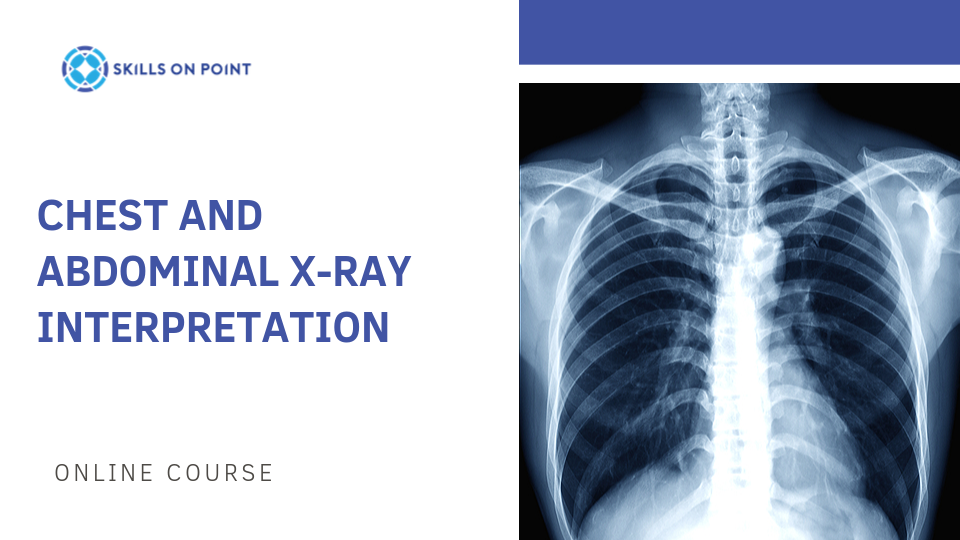
One question I constantly get asked as a nurse practitioner is how to get comfortable and confident at x-ray interpretation and identifying chest X-ray findings. The best advice is to be consistent with your approach of evaluating the images you see every single day and as you get used to seeing normal findings, abnormal findings will jump out at you. Here is the way I recommend you evaluate the chest X-ray to avoid missing subtle findings.
Tips for Chest X-Ray Interpretation
Step 1: Know Your Resources
Especially when starting out, have an expert to help you go through the image. This may be and on call radiologist, or it may be a peer health care provider with more experience than you.
Step 2: Use the Alphabet Method
Airway: Look for the location and visibility/patency of the trachea and carina. This step is also helpful for identifying if your images properly aligned or off-axis.
Bones/Breast: Evaluate the cortex of the bones to see if there are any evidence of fracture, healing fracture, and overall exposure of the image. breast tissue may or may not be present, as well as piercings, implants, and other external extras. It’s important to recognize that although the breast tissue looks inferior on the image, that is an optical illusion. The reality is that the chest X ray is much more superior in the chest then we may think it would be and the inframammary creases are totally normal.
Cardiac Silhouette: Inspect for size, location, and any obliteration of the cardiac margin by adjacent structures. many people who have pneumonia will have an obliteration of the cardiac silhouette known as “silhouette sign”. this helps direct the interpreter of the image as to which lobe of the lung has the pneumonia. an enlarged heart may suggest cardiomegaly, hypertrophy, dilated cardiomyopathy, or other pericardial pathology like tamponade.
Diaphragm: Identify the diaphragm bilaterally and no whether one side is higher than the other, or if there is evidence of free air inferior to the diaphragm. Unless a recent abdominal surgery was performed, any evidence of free air should be considered a emergency as it may likely reflect perforated gastric ulcer.
Extrathoracic Tissue: Consider the size of the bone structure compared to the extra thoracic tissue. If your patient has COPD, expect to see of minimal amount of extra thoracic tissue, or if the patient has considerable obesity in the upper torso, expect to find a smaller bony frame of the patient in relationship to their total body mass and skin. You might also appreciate subcutaneous air or any traumatic impalement and the extra thoracic tissue.
Fields of the Lung: Evaluate whenever possible both anterior/posterior and lateral imaging to evaluate which lobe may be involved in a consolidation, atelectasis, pneumonia, or nodule. one nice trick for evaluating lobes is to remember that the upper lobes are much like shingles on your roof and they overlap the lower lobes. the right middle lobe and the lingula of the left upper lobe are much like two hands reaching forward holding the heart. If you notice obliteration of the cardiac margin, aka “positive silhouette sign”, this would be indicative of a right middle lobe or a left upper lobe lingula consolidation. Also evaluate for evidence of Kerley B lines to suggest layering of interstitial fluid and edema. This may indicate fluid overload or a cardiogenic etiology such as systolic dysfunction.
Gastric Bubble: Evaluate the gastric bubble for its presence, and also its absence. Patients who are supine during imaging will likely not have a gastric bubble present since the some of the contents will layer out against the plate and uniformly be viewing through the gastric bubble, but for patients who are sitting erect, a gastric bubble should be visible. this is also helpful for identifying feeding tubes and identifying the stomach landmarks for the fundus, antrum, lower esophageal sphincter, and outflow into the duodenum.
Hilum/Mediastinum: Inspect the hilum for evidence of enlarged lymph nodes, masses, overlying lung involvement, and pulmonary edema. also evaluate the mediastinal AM for evidence of aortic enlargement, calcified vasculature, masses, and lymphadenopathy.
Indwelling Lines/Instrumentation: For hospitalized patients, make sure to include an evaluation for any indwelling lines or tubes used actively in the patients acute care management such as endotracheal tubes, surgical drains, chest tubes, feeding tubes, central lines, peripherally inserted central lines, balloon pumps, ventricular assist devices, and any other imaginable device. For patients who are outpatient, expect to find things such as pleural catheters, ports, peripherally inserted central lines, and spinal hardware.
Step 3: Clinical Correlation is a Must
Sometimes radiologists will use the term “clinical correlation is required”. this is a very important statement and it’s not the radiologist passing the buck to the provider or simply raising their hands in the air and saying “I dunno”. Much more importantly than any medical image is your job as a health care provider to put your hands on the patient in evaluation, and to make sound medical decision making based off of the patient’s history and presenting signs/symptoms.
Medical imaging is only as useful as the provider requesting the image to give a clear and concise expectation of what the concerns are that the radiologist should be looking for in their evaluation. sometimes a phone call before the image saves the patient from receiving an inappropriate image modality and spending a few extra seconds to write out what you’re looking for helps the radiologist clue in on what you’re hoping to rule in or rule out. remember, the radiologist is not at the bedside and doesn’t see the patient. They are trained to evaluate what it could be or what it couldn’t be based off of the image, and the image alone.
Step 4: Serial Imaging Should be Viewed Whenever Possible
The radiologist performing the x-ray interpretation of the image will most certainly look into the system to find additional images if there is anything in question, and I will be the first one to admit that chest X rays are very simple, quick look. Some pathology requires a more advanced image such as a chest CT, angiography, or MRI. If those have been performed, the radiologist can correlate existing findings compared to those other more advanced findings, which is why I absolutely recommend you talk to your radiologists and be specific in seeking x-ray interpretation training to make sure you know what you’re looking at when you do evaluation.
I hope this is useful to get your mind thinking about how to successfully navigate the chest x-ray. If you would like to learn more about this, check out our course: Chest and Abdominal X-Ray Interpretation and all the other nursing continuing education courses on our online CE portal at: skills-on-point.teachable.com


